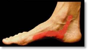
Posterior Tibial Tendonitis
Posterior Tibial Tendonitis also know as Adult Acquired Flatfoot Deformity is a very common problem. The process can occur in nearly anyone, but it is most commonly seen in middle age or elderly females. The cause is multifactorial but has been linked to hypertension, diabetes, obesity, and many other contributing factors.
Early in the disease process non-operative treatment may help relieve pain and slow progression. However, once a deformity develops and begins to rapidly progress, surgery may be necessary to realign the foot and prohibit further collapse.

The primary cause begins with dysfunction of the posterior tibial tendon which normally functions to maintain the arch of the foot. With continued posterior tibial tendon degeneration, the arch can collapse leading to a flat foot deformity. The final product of the disease process is often the result of several mitigating abnormalities throughout the foot which develop in time as normal posterior tibial tendon function is lost.
Development of deformity in multiple anatomic planes leads to a dysfunctional and maligned foot and ankle. Most commonly the deformity includes forefoot varus, midfoot abductus, hindfoot valgus, and ankle valgus. These are simply descriptive terms use by surgeons to help characterize the different components of the malalignment. We mention these terms only because it is important for you to understand that sometimes multiple incisions and procedures are needed to help separately address each of the components of malalignment.
The disease process has been categorized into four stages based on progression of the deformity and surgical management can be tailored depending on the your classification stage:
Stage I- Tendon Pain and Dysfunction Without Deformity
Stage II- Flexible Flatfoot Deformity
Stage III- Rigid Flatfoot Deformity
Stage IV- Flatfoot Deformity with Progression to Ankle Degeneration
Regardless of the stage of adult acquired flatfoot deformity, conservative treatment may be beneficial. In early stages pain relief may be obtained by resting the tendon to allow inflammation resolution followed by physical therapy to strengthen the tendon. The tendon may be rested with activity modification and also by relieving tension on the tendon with casting, booting, bracing, or arch supports. Oral anti-inflammatory medications and occasionally a steroid injection may be beneficial. When conservative treatment fails, then operative intervention can be very beneficial.
Stage I is generally characterized by a painful posterior tibial tendon that is still functional. Stage I can often be treated with debridement of the inflamed tendon or tendon sheath.
Stage II is characterized by dysfunction of the posterior tendon in which a flatfoot deformity develops as the arch begins to collapse. The foot remains flexible, and therefore, the dysfunctional posterior tibial tendon can be resected and another tendon (usually the flexor digitorum longus tendon) can be transferred into its position to restore function. It may be necessary to perform simultaneous balancing procedures to the foot to protect the transferred tendon and to maintain restoration of the arch. These additional procedures are tailored to each patient but may include a gastrocnemius lengthening, a lateral column lengthening, a Cotton osteotomy, a medial slide calcaneal osteotomy, or selective fusion of certain joints.
Stage III describes a situation in which the tendon dysfunction leads to a rigid flatfoot deformity that cannot be passively corrected. In Stage III, it is often necessary to fuse multiple degenerative joints in the foot to restore alignment and to protect the ankle joint from degenerative arthritis. These selective fusions may be done in combination with additional procedures used in earlier stages of the deformity.
Stage IV is characterized by many of the same issues as earlier stages, but the distinguishing factor is that the ankle joint becomes involved. A neglected flatfoot deformity leads to an imbalance of forces at the ankle joint which can progress to degenerative arthritis with erosion of the ankle joint cartilage. A Stage IV deformity is treated similarly to other stages of the deformity with selective fusions and balancing procedures in the foot. However, the ankle joint generally needs to be addressed as well. Typically, the ankle joint is treated with a fusion or with a resurfacing utilizing a total ankle replacement. Sometimes a Stage IV deformity may require one operation to address the foot and a second operation to address the ankle.
The recovery process from flatfoot surgery varies depending on which stage of posterior tibial tendonitis you have and what procedures are necessary to correct the deformity. Much of the time immediately after surgery is spent resting and elevating your leg to decrease swelling. We generally recommend you keep your leg elevated as much as possible during the first week. You will be discharged from the hospital immediately after surgery or on post-op day 1 with a prescription for pain medicine to help control your pain as the nerve block begins to wear off. Most patients are discharged home in a splint which will be converted to a cast in the office. Stitches are generally removed around 2-3 weeks. Most patients are placed in a postoperative non-weight bearing cast for six weeks followed by a walking cast for another six weeks. You will not be able to put weight on your ankle during the first six weeks and will need to use crutches, a walker, or a rolling knee walker to mobilize. Patients are generally transitioned from a cast to a postoperative walking boot at about week 12 and then into a regular shoe. Physical therapy may be beneficial to regain strength and motion. The total recovery process may take up to six months.